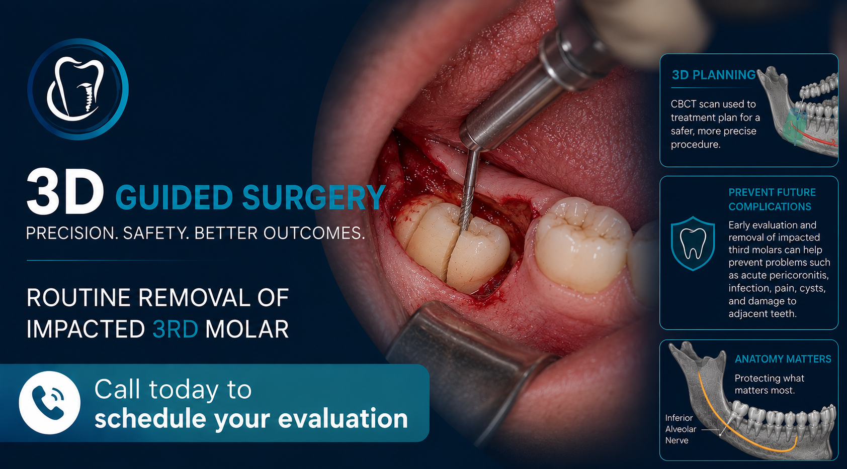
It is important for patients who show signs of nerve compression or nerve deviation (known as Rood’s criteria) on their panoramic image (2D image) to be well informed about the need of a dental CBCT (3D image). We recommend this to give them the best opportunity to avoid damage to the mandibular neurovascular bundle also known as the inferior alveolar nerve (IAN) peri-operatively. Damage to the mandibular IAN can present as temporary or permanent numbness and/or tingling of the lower lip or chin. The risk of permanent nerve damage occurring is usually low (around 1%), however sometimes the patient’s anatomy may indicate they have increased risk. We do our best to identify signs of increased risk on 2D imaging (signs of nerve compression, deviation, or entrapment) and suggest the CBCT imaging when we feel it is indicated.
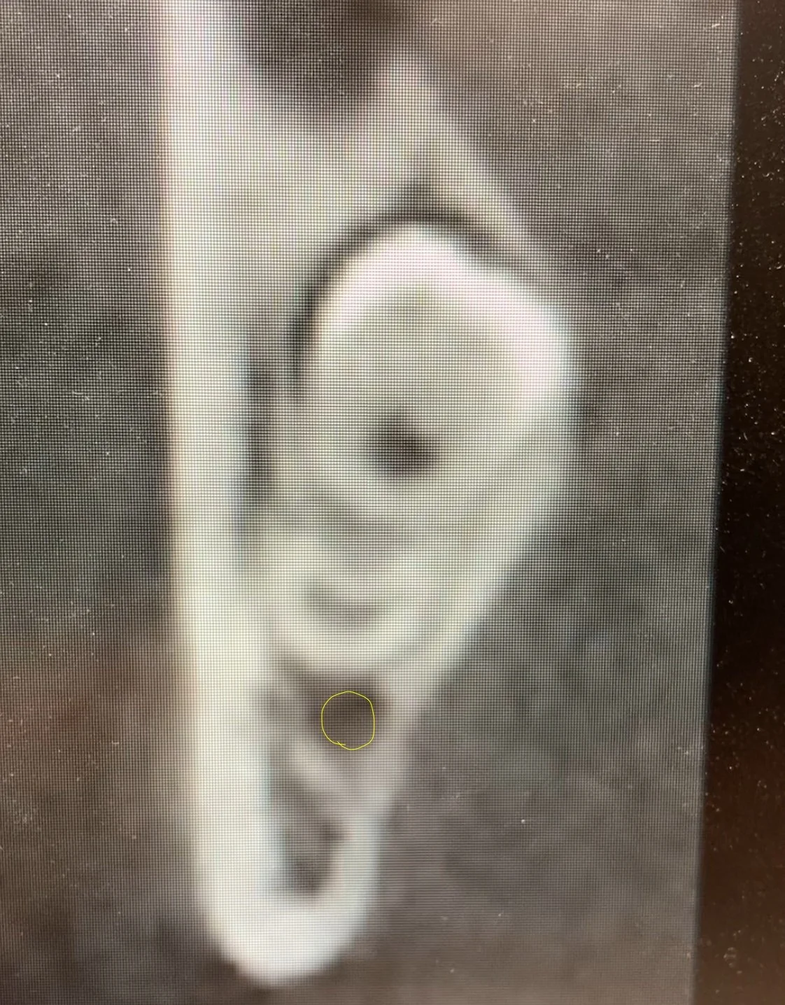
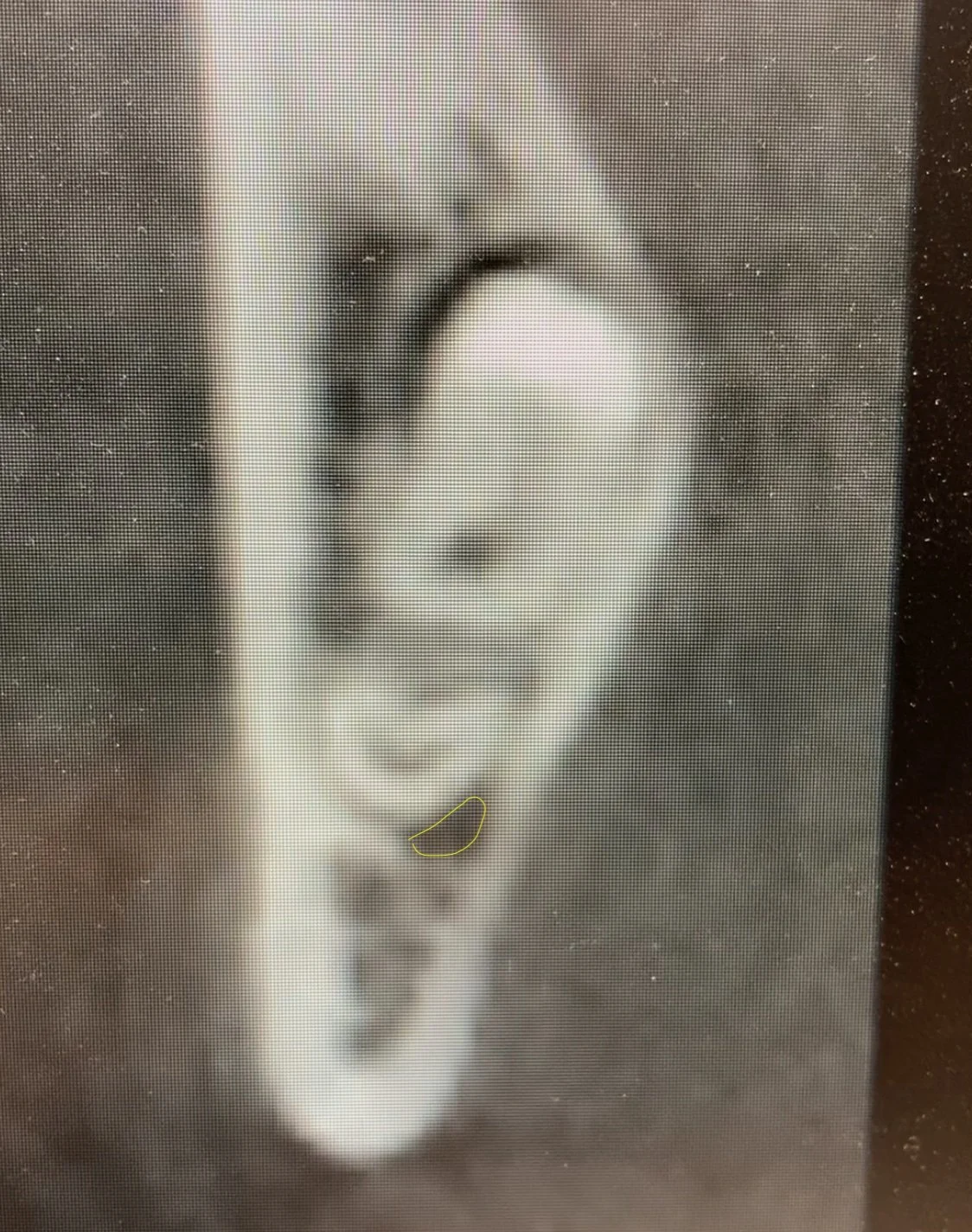
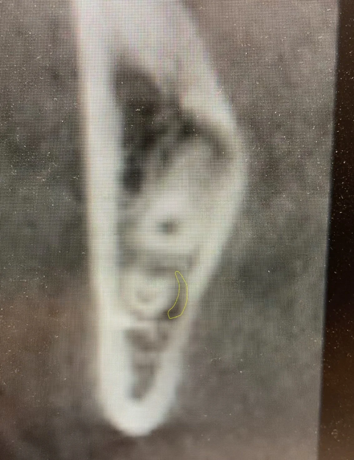
Notice how the nerve canal changes shape as it traverses this portion of mesial root surface of an impacted third molar. This is a risk factor which corresponds to a significant increase in the risk of damage to the IAN when the tooth is removed. This kind of risk factor can only be fully evaluated prior to the surgical procedure by using 3D imaging such as a dental CBCT.
Treatment such as a coronectomy may be recommended in cases where risks are determined to be increased.
Note:
There is always some risk associated with surgical procedures, and patients must make their own decision regarding treatment vs. no treatment, considering the pros and cons of each option.
CBCT Imaging is not always indicated for extractions, any recommendation will be made based on existing radiographs (E.g. Panoramic radiograph).