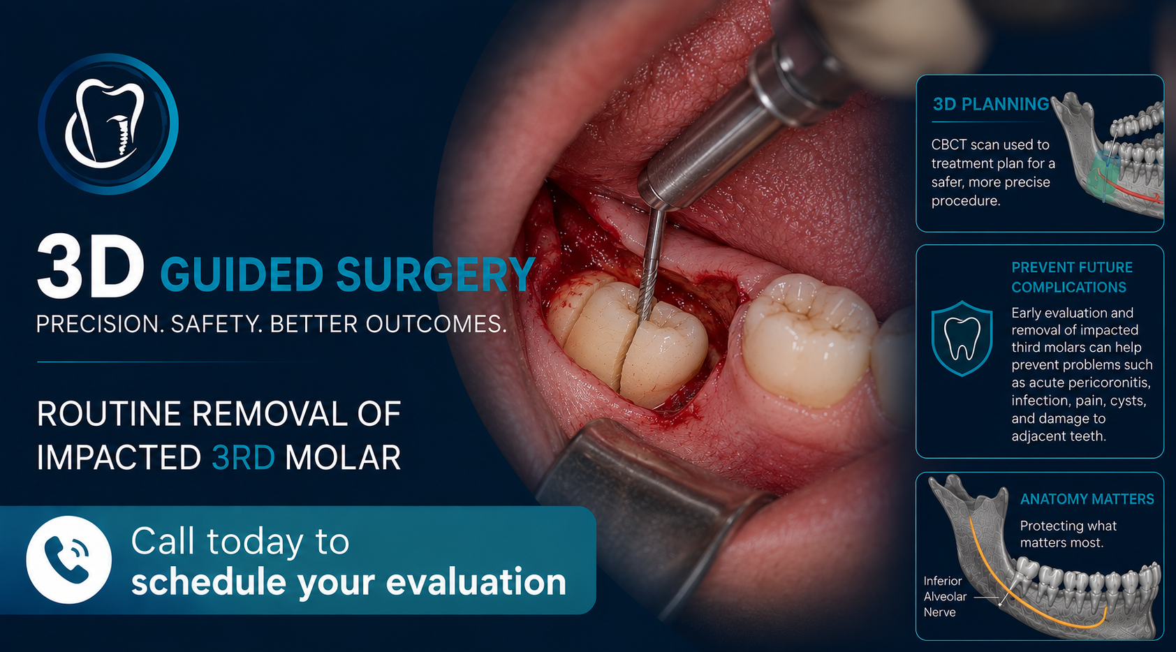
It is important for patients who show signs of nerve compression or nerve deviation (known as Rood’s criteria) on their panoramic image (2D image) to be well informed about the need of a dental CBCT (3D image). We recommend this to give them the best opportunity to avoid damage to the mandibular neurovascular bundle also known as the inferior alveolar nerve (IAN) peri-operatively. Damage to the mandibular IAN can present as temporary or permanent numbness and/or tingling of the lower lip or chin. The risk of permanent nerve damage occurring is usually low (around 1%), however sometimes the patient’s anatomy may expose them to increased risk. We do our best to identify signs of increased risk on 2D imaging (signs of nerve compression, deviation, or entrapment) and suggest the CBCT imaging when we feel it is indicated. Treatment such as a coronectomy may be recommended in cases where risks are determined to be increased.
CBCT imaging can also be useful for other impacted teeth, such as canines, and when treatment planning expose and bond procedures. CBCT imaging is becoming the standard of care for root canal treatment to avoid missing anatomy (E.g. extra canals which may be hidden on traditional PA radiographs).
CBCT Imaging is not always indicated for extractions, any recommendation will be made based on existing radiographs (E.g. Panoramic radiograph).